
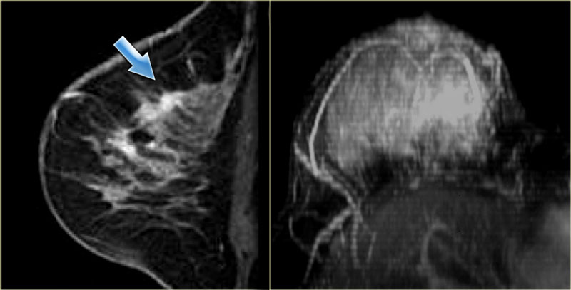

The image on the left shows an enhancing mass in the left breast. Patients were meticulously scanned with both grey scale and Doppler US. Tozaki et al. LEFT: Fibroadenoma with non-enhancing septations. This was in agreement with Lobbes et al who compared CESM versus MRI for the assessment of the size of the breast tumor; they found that CESM is good as an imaging tool for measuring the size of the tumor and MRI did not improve the quality of the tumor size evaluation [ 20 ]. An ultrasound guided core biopsy was done and histopathologically proven as granulomatous mastitis. On ultrasound b , the lesion appears as a fairly defined hypoechoic lesion with a tongue-like projections arrows.


If the lesion was identified, then the skin area was ink-marked and sterilized. Also, low-energy images of CESM similar to digital mammography are able to detect micro-calcifications, speculated lesion, and architectural distortion which may be missed on subtracted CESM and MRI if not enhancing [ 10 ]. Patients were meticulously scanned with both grey scale and Doppler US. Then there is the delayed portion - two minutes or more after the injection of contrast. In conclusion although in our study Washout pattern was the most powerful indicator for malignant pathology in non mass like enhancing lesions, more studies with larger sample size needs in this regard. Received: December 13,